A couple weeks ago, I introduced the Rapid Fluoroscopy application and hinted that it could be used for more than the typical fluoroscopy applications. In this post, I’m going to elaborate on that.
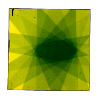
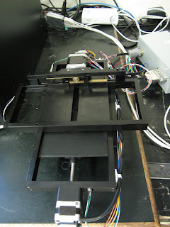
Parallax Innovations has been working with Dr. Eugene Wong and Ph.D. candidate Michael Jensen at the University of Western Ontario, who have customized their eXplore CT120 to allow it to be used for image-guided radiation delivery. In particular, they have added a motorized, computer controlled collimator (shown below) to the system that allows them to narrow the x-ray beam for targeting a specific region of the specimen. The image to the right is of a 4x4 cm radiochromic film showing how they can achieve an elliptical radiation pattern.
The problem that we helped them solve was how to define a series of exposures at prescribed angles as part of a micro CT protocol. They needed the ability to fire any number of exposures with any allowable x-ray settings from any gantry position. And in addition, they needed to be able to control the size of the collimator aperture at any given gantry position. Except for the control of custom collimator, the requirements seemed to me to be close to the functionality already provided by the Rapid Fluoroscopy application. Parallax Innovations assisted by implementing a new custom protocol to the application – the treatment protocol.
Treatment protocols allow the operator to specify any number of gantry angles and for each angle, specify a number of other properties. As you can see in the property editor pictured below, one can prescribe the x-ray settings (voltage/current/duration), a table offset from the landmark position, the focal spot size, and how many exposures to fire. There are also settings for the maximum allowable anode heat before the exposures at this particular position can begin (anode heat threshold), the amount of time between exposures (decay time), and an option to wait for a given amount of time before beginning the exposures at this particular position (pause).
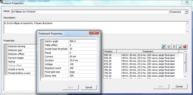
Note: the 140 kV voltage setting is not available with standard x-ray generators. 120 kV is normally the maximum.
For the custom collimator, Michael had already written an application to control it. I asked him to add a remote procedure call (RPC) interface that would allow the fluoroscopy software to make RPC calls passing the collimator motor positions defined in the treatment protocol.
A custom protocol using Rapid Fluoroscopy was implemented and integrated with collimator control in a timely and efficient manner. It provided a flexible and simple solution which accelerated the readiness of such a platform for targetted radiation delivery. Now, when the Rapid Fluoroscopy software is switched on, it is possible to build and run radiation treatment protocols. Such a combination can also be used for cardiac imaging, with the collimator shielding the rest of the animal from unnecessary radiation exposures, which could potentially improve image quality by reducing scattered radiation. For more information about Dr. Wong’s research, visit the University of Western Ontario website .
Special thanks to Michael Jensen for all the testing he has done with the treatment protocols.
Comments